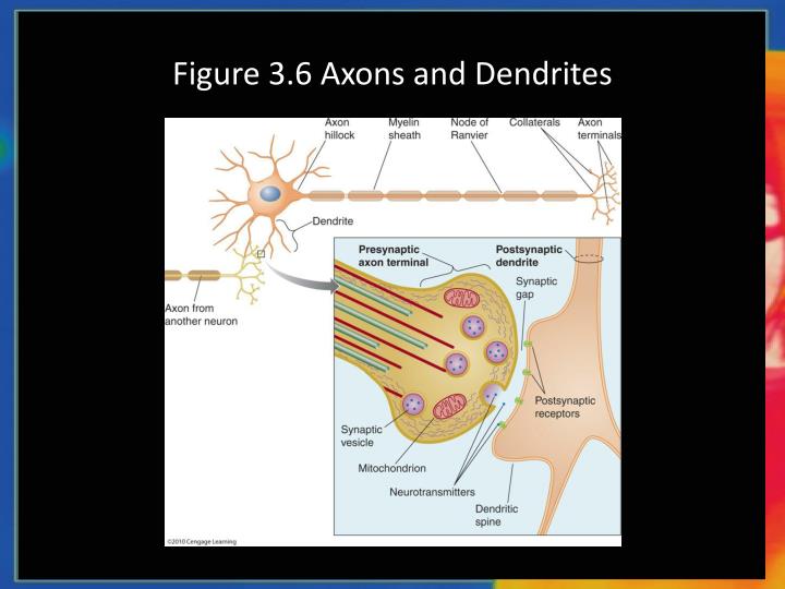

The cookie is used to store the user consent for the cookies in the category "Performance". In our theoretical analysis, we have assumed that the AIS diameter d is fixed, but it is possible to take diameter changes into account in our analysis. This cookie is set by GDPR Cookie Consent plugin. The axon-carrying dendrite starts with diameter 2 µm from the soma and splits after 7 µm into two branches of equal diameter (such that d main 3 / 2 d 1 3 / 2 + d 2 3 / 2). The cookie is used to store the user consent for the cookies in the category "Other. This cookie is set by GDPR Cookie Consent plugin. The cookies is used to store the user consent for the cookies in the category "Necessary". The cookie is set by GDPR cookie consent to record the user consent for the cookies in the category "Functional". The cookie is used to store the user consent for the cookies in the category "Analytics". Careful examination of the morphological transition between neural progenitors and post-mitotic neurons reveal. While migrating, post-mitotic neurons form a leading process and a trailing process which become the axon or the dendrite depending on the cell type (Figure 1). This cookie is set by GDPR Cookie Consent plugin. In vivo, most neurons undergo axon-dendrite polarization during migration. These cookies ensure basic functionalities and security features of the website, anonymously. Necessary cookies are absolutely essential for the website to function properly. There are not such vesicles in the dendrites.Īxon contains neurofibrils all over but they lack Nissl’s granules.ĭendrites contain both neurofibrils and Nissl’s granules.Īxon conducts neuronal impulse away from the soma (cell body).ĭendrites conduct neuronal impulses towards the soma. No synaptic knots are formed on the tip of the dendrites. The terminal branches f the axon forms an enlarged synaptic knot. The thickness of axon is uniform throughout the length.ĭendrites are highly branched throughout their length. (Image Source: CC Wikipedia) You may also like NOTES in.Īxon arises from the discharging end of the nerve.ĭendrites arise from the receiving surface of the nerve.Īxon is comparatively long, sometimes several meters.ĭendrites are comparatively shorter (usually under 1.5 mm) Axons have tau-bound microtubules of uniform orientation, whereas dendrites have. Ø Both are cytoplasmic projections from the cell body of a neuronal cell. Microtubule organization and organelle distribution in axons and dendrites. Ø Both axon and dendrites are the parts of neuron.


 0 kommentar(er)
